
What are Glycoconjugates?
Polysaccharides and oligosaccharides act also as information carrier molecules. Informational carbohydrate is covalently joined to a protein or lipid to form glycoconjugates. As the name suggests, the glycoconjugates are conjugate (joined) molecules of carbohydrates other macromolecules such as proteins or lipids. The present post discusses the structure, classification and functions of important glycoconjugates.
Table of Contents
@. Functions of Glycoconjugates
@. Examples of Glycoconjugates
$. Proteoglycans
$. Glycoproteins
$. Glycolipids & LPS
@. Proteoglycan vs Glycoprotein
@. Related Posts
Functions of Glycoconjugates
Ø Glycoconjugates acts as information carrier molecules.
Ø They serve as destination labels for some proteins.
Ø They serve as mediators of specific cell-cell interaction.
Ø They can act as the mediators of interactions between cell and ECM.
Ø They form Glycocalyx (Glycocalyx is a carbohydrate layer formed by specific oligosaccharide chains attached to components of the plasma membrane).
Ø Specific carbohydrate-containing molecules from the glycocalyx help in:
$. Cell-cell recognition
$. Adhesion
$. Cell migration during development
$. Blood clotting
$. Immune response
$. Wound healing
| You may also like NOTES in... | ||
|---|---|---|
| BOTANY | BIOCHEMISTRY | MOL. BIOLOGY |
| ZOOLOGY | MICROBIOLOGY | BIOSTATISTICS |
| ECOLOGY | IMMUNOLOGY | BIOTECHNOLOGY |
| GENETICS | EMBRYOLOGY | PHYSIOLOGY |
| EVOLUTION | BIOPHYSICS | BIOINFORMATICS |
Examples of Glycoconjugates
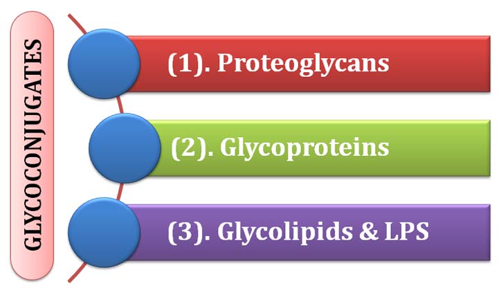
Ø There are THREE important types of glycoconjugates in the cells. They are:
(1). Proteoglycans
(2). Glycoproteins
(3). Glycolipids and Lipopolysaccharides (LPS)
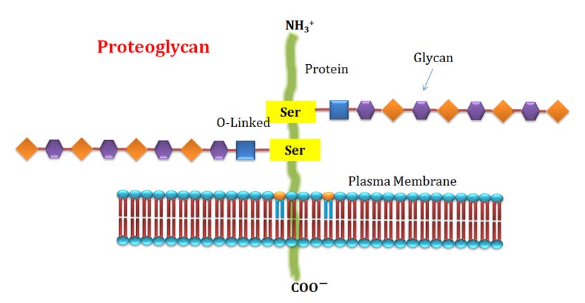
(1). Proteoglycan
Ø In proteoglycan, one or more sulfated glycosaminoglycan chains are joined covalently to a membrane protein or a secreted protein.
Ø They are macromolecules of cell surface and extracellular matrix.
Ø Glycan moiety commonly forms the greater fraction by mass.
Ø In proteoglycan, glycan forms the main part of biological activity.
Ø Glycan part provides provisions for hydrogen bonding due to the presence of many – OH groups.
Ø Glycan part provides electrostatic interactions (due to the presence of sulfate groups in glycosaminoglycans) with other proteins of ECM.
Ø Proteoglycans are major components of connective tissue such as cartilage.
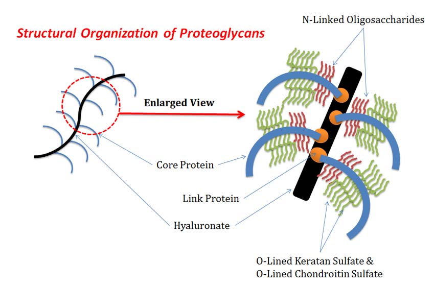
Structural Organization of Proteoglycans
Ø Proteoglycans have a bottlebrush-like molecular architecture with ‘bristles’.
Ø ‘Bristles’ are attached non-covalently to a filamentous hyaluronate backbone.
Ø The ‘bristles’ consist of a core protein to which glycosaminoglycans (GAGs) are covalently linked.
Ø The GAGs are most often chains of keratan sulfate and chondroitin sulfate.
Ø Interaction between core protein and hyaluronate is stabilized by a link protein.
Ø Smaller oligosaccharides are usually attached to the core protein near its site of attachment to hyaluronate.
Ø These oligosaccharides are glycosidically linked to the protein via the amide N of specific Asparagine residues (and are therefore known as N-linked oligosaccharides).
Ø The keratan sulfate and chondroitin sulfate chains are glycosidically linked to the core protein via oligosaccharides that are covalently bonded to the side-chain O atoms of specific Serine or Threonine residues (i.e., O-linked oligosaccharides).
Types of Proteoglycans
Ø Proteoglycans are categorized based on two criterion.
(1). Based on their relative size: Large Proteioglycans and Small Proteoglycans
(2). Based on the type of their glycosaminoglycan chains
Ø Based on the glycosaminoglycan chain, there are three categories of proteoglycans:
$. Heparan sulfate proteoglycan (HSPGs)
$. Chondroitin sulfate proteoglycan (CSPGs)
$. Keratan sulfate proteoglycan (KSPGs)
(2). Glycoprotein
Ø Glycoproteins have one or several oligosaccharides of varying complexity joined covalently to proteins.
Ø Usually the protein part in glycoprotein will be the bulky part when compared to proteoglycan.
Ø Glycoproteins are found on the:
$. Outer surface of plasma membrane
$. In the extracellular matrix
$. In the blood
Ø Inside the cells they are found in specific cell organelles such as Golgi complex, Secretory granules and Lysosomes.
Ø Carbohydrate part is attached to proteins through its anomeric carbon.

Ø The anomeric carbon attached to the protein through:
(a). An O-glycosidic link to the —OH of a Ser or Thr residue (O-linked)
(a). An N-glycosidic link to the amide nitrogen of an Asn residue (N-linked)
Ø Most of the glycoproteins contain many carbohydrate units.
Ø The carbohydrate content of glycoprotein range from 1% to 70% of the total mass.
Ø Some glycoproteins have a single oligosaccharide chain.
Ø About half of all proteins of mammals are glycosylated.
Functions of Glycoproteins
Ø The glycan part in the glycoprotein carries rich information.
Ø They form highly specific sites for recognition, binding and interactions for other proteins.
Ø Glycoproteins are important for the recognition of white blood cell.
Ø Glycoproteins are also involved in the immune system of mammals.
Ø Antibodies (immunoglobulins), which directly with antigens contain are glycoproteins.
Ø Many molecules of MHC (Major Histocompatibility Complex) are glycosylated proteins belong to glycoprotein category.
Ø The Sialyl Lewis X antigen on the surface of leukocytes is glycoproteins.
Ø The H antigen of the ABO blood is a glycoprotein.
Ø The follicle-stimulating hormone gonadotropin is a glycoprotein.
Ø The zona pellucida of ovum contains many glycoproteins, which helps in the recognition and interaction between egg and sperm cells.
Ø GP-120 (Glycoprotein-120) and GP-41 (Glycoprotein-41) are HIV viral coat proteins.
Ø Egg white and blood plasma proteins are mainly glycoproteins, they form viscous fluids.
Glycoprotein vs Proteoglycan:
Ø In glycoproteins the carbohydrate portion is smaller than that in proteoglycan.
Ø In glycoproteins the carbohydrate moiety shows more structural diversity than proteoglycan.
(3a). Glycolipids
Learn more: Membrane Lipids
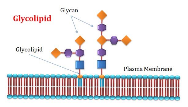
Ø They are membrane lipids in which the hydrophilic head groups are oligosaccharides.
Ø Glycerolipids and Sphingolipids are the two important categories of glycolipids which have glycerol or sphingosine backbones respectively.
Ø Similar to glycoprotein, glycolipids also act as specific sites for recognition by carbohydrate-binding proteins.
Ø The gangliosides (a heavily carbohydrate attached membrane lipid) are membrane lipids of eukaryotic cells.
Ø In gangliosides the polar head group is a complex oligosaccharide chain with one or many sialic acid residues.
Ø Oligosaccharide groups of gangliosides determine the ABO blood group in human.
| You may also like... | ||
|---|---|---|
| NOTES | QUESTION BANK | COMPETITIVE EXAMS. |
| PPTs | UNIVERSITY EXAMS | DIFFERENCE BETWEEN.. |
| MCQs | PLUS ONE BIOLOGY | NEWS & JOBS |
| MOCK TESTS | PLUS TWO BIOLOGY | PRACTICAL |
(3b). Lipopolysaccharides (LPS)
Ø Lipopolysaccharides (LPS) are found in the outer membrane of Gram-negative bacteria (Eg. E. coli).
Ø LPS is a very complex macro-molecule of lipid and carbohydrates.
Ø Different bacteria have different structure of LPS, but they have a common structural backbone.

Source: wikipedia
Ø All LPS have a common structure of:
§ Fatty acid residues (Called Lipid-A, or Endotoxin)
§ A core oligosaccharide
§ an “O-specific” chain (O-antigen)
Ø The O-antigen the principal determinant of the serotype of the bacterium.
Ø LPS is responsible for the production of antibodies by the animal immune system as a response to microbial infection.
Ø LPS provoke Tol-like-receptor (TLR) and promote inflammation in mammalian cells.
Ø The outer membranes of the gram-negative bacteria S. typhimurium and E. coli contain so many lipopolysaccharide molecules that the cell surface is virtually covered with O-specific chains.
References
@. Lehninger A.B., (2018), Textbook of Biochemistry, Ed. 5, Pearson International, New York
@. Voet, D., Voet, J.G. and Pratt, C.W., 2013. Fundamentals of biochemistry: life at the molecular level
Do you have any Queries? Please leave me in the Comments Section below.
I will be Happy to Read your Comments and Reply.
<<< Back to Biochemistry Notes Page
Introduction to Carbohydrates | Monosaccharides | Disaccharides | Polysaccharides | Glycosaminoglycans | Sugar Derivatives | Glycoconjugates |
More Lecture Notes from Easy Biology Class…
BotanyZoologyBiochemistryGeneticsCell & Molecular BiologyBiotechnologyPhysiology & EndocrinologyPlant PhysiologyMicrobiologyImmunologyEmbryologyEcologyEvolutionBiophysicsResearch MethodologyBiostatisticsChemistry for BiologistsPhysics for Biologists
Browse more in Easy Biology Class…
Lecture NotesBiology PPTsVideo TutorialsBiology MCQQuestion BankDifference betweenPractical AidsMock Tests (Online)Biology Exams