Hip Anatomy
The hips and structures within the hips are some of the most important and complex structures in the human body. They play a crucial role in our everyday movements, from walking and running to sitting and standing. Understanding Hip Anatomy is essential to maintaining optimal hip health and longevity. Join us as we explore the fascinating intricacies of the hips, from the bones and joints, to the muscles, ligaments and primary functions that support our everyday movements.
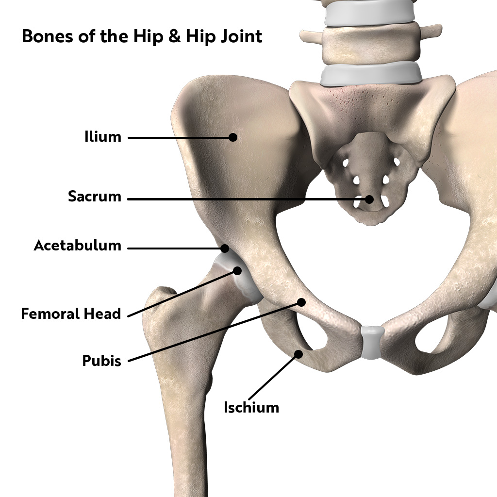
Bones of the Hip
There are five bone structures that make up the hip. Three of which come together to form the primary hip structures: the Ilium, the Ischium, and the Pubis, with the other two, the Femoral Head and the Acetabulum, forming the hip joint.
- Ilium: This large, wing-shaped bone forms the upper part of the hip, providing stability and support for the joint.
- Ischium: The ischium is the lower and posterior part of the hip bone. It forms the “sit bones” and supports our body weight when we sit.
- Pubis: The pubis is the anterior part of the hip bone and is responsible for connecting the two hip bones at the front of the pelvis.
The Hip Joint
The hip joint is a ball-and-socket joint, designed for both mobility and stability. It consists of two primary components:
- Femoral Head: The rounded head of the femur (thigh bone) fits into the acetabulum, creating the ball part of the ball-and-socket joint. This structure allows for a wide range of motion in multiple directions.
- Acetabulum: The acetabulum is the socket or cup-shaped structure in the hip bone that receives the femoral head and provides stability to the joint.
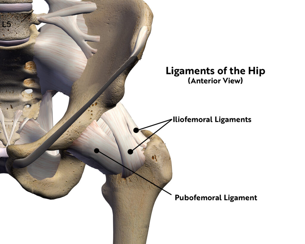
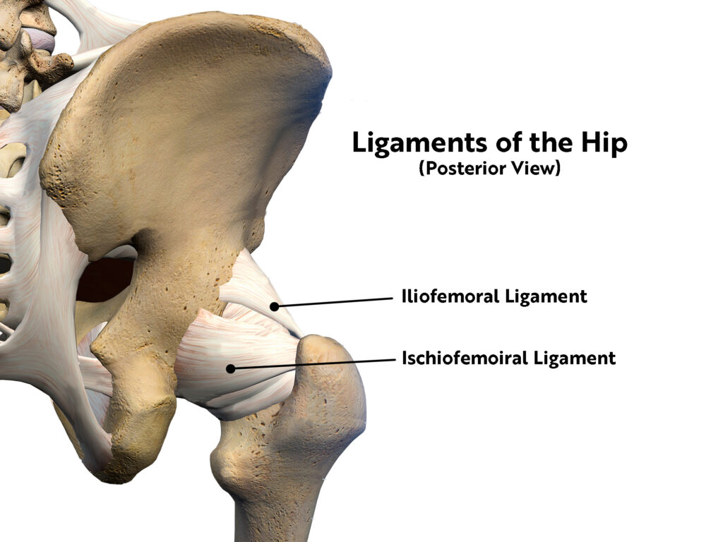
Ligaments of the Hip
Within our hip anatomy, we have several ligaments that hold the bones of the hip together and provide stability to the joint.
- Iliofemoral Ligament (Y-shaped ligament): This ligament is the strongest in the body and helps to prevent overextension of the hip joint.
- Pubofemoral Ligament: This ligament reinforces the front of the hip joint and assists in preventing hyperextension.
- Ischiofemoral Ligament: Located on the back of the hip joint, this ligament stabilizes the joint during internal rotation and extension.
Muscles of the Hip
Now that we’ve covered the bones and ligaments that form the hips’ basic structures, let’s dive into the anatomical components that give us the strength and support to move and stabilize ourselves through space: the muscles of the hip.
The hip joint is surrounded by a complex network of muscles. Some of the key muscles include:
- Gluteus Maximus: This is the largest and most powerful hip muscle, responsible for hip extension (extending the leg backwards) and external rotation (moving the leg outwards).
- Gluteus Medius and Minimus: These muscles lie on the outer side of the hip and are responsible for hip abduction (moving the leg away from the body), internal rotation (rotating the leg inwards) and external rotation (rotating the leg outwards).
- Iliopsoas: This is a group of muscles responsible for hip flexion (bending the hip joint), helping you lift your knee towards your chest or hinging over to pick something up.
- Adductors: The adductor muscles on the inner thigh allow for hip adduction (moving the leg toward the midline of the body).
- Iliotibial Band (IT Band) is a thick band of muscle fascia that originates at the lateral portion of the iliac crest and inserts at the lateral condyle of the tibia (shin bone). Its main function is the provide stabilization to the pelvis and aid in posture control.


Movements of the Hip
These muscles help facilitate the four primary movements of the hip: Flexion, Extension, Internal Rotation, and External Rotation.
- Hip Flexion: involves lifting the thigh toward the abdomen or vice versa, bringing the abdomen closer to the thigh. This movement is essential for activities like walking, running, lifting the leg, walking up stairs, hinging over to pick something up, sitting in a chair, etc.
- Hip Extension: involves extending the leg backward and is executed when your back leg is straightened as you walk. You execute hip extension anytime you stand up from a seated position or run.
- Hip Internal Rotation: occurs when the thigh is rotated or turned inwards towards the midline of the body. Anytime we cross our legs, we are performing hip internal rotation.
- Hip External Rotation: occurs when the thigh is rotated or tuned outwards, away from the body. We perform this movement when we take a turn during a walk or sit in the butterfly stretch position during a yoga class.
It’s no wonder, when we injure even a small structure in our hips, it can take its toll on our everyday movements. This is why, we at Colorado Springs Orthopaedic Group emphasize strengthening your hip muscles, core, and leg muscles in efforts of preventing injury during activity. Strengthening these muscles through a full range of motion will also aid in maintaining your mobility.
Whether you’re an athlete or just someone who values their joint health, be sure to check out our Healthy Hips series on our website at https://www.csog.net/blog/ to learn more about hip anatomy. Tune into our Youtube channel to learn how taking an interest in ways to optimize hip strength and mobility can dramatically affect your hip and overall physical longevity.